


Popular Articles
- Earliest molecular events of vision revealed
- Dynamics and Kinetics in Structural Biology
- XFEL Pulses Demonstrate How Plants Perceive Light
- Structural biology is solved -- now what?
- BioXFEL Postdoctoral Fellowship Award
Archived Articles
- Details
- Tuesday, 02 May 2017
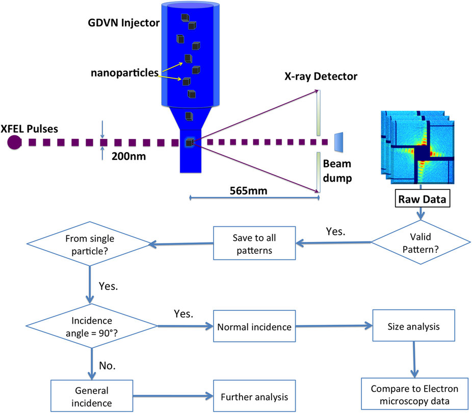
X-ray free-electron lasers provide novel opportunities to conduct single particle analysis on nanoscale particles. Coherent diffractive imaging experiments were performed at the Linac Coherent Light Source (LCLS), SLAC National Laboratory, exposing single inorganic core-shell nanoparticles to femtosecond hard-X-ray pulses.
Each facetted nanoparticle consisted of a crystalline gold core and a differently shaped palladium shell. Scattered intensities were observed up to about 7 nm resolution. Analysis of the scattering patterns revealed the size distribution of the samples, which is consistent with that obtained from direct real-space imaging by electron microscopy. Scattering patterns resulting from single particles were selected and compiled into a dataset which can be valuable for algorithm developments in single particle scattering research.


